Doppler Ultrasound During Pregnancy
Contents:
- Doppler Sonography During Pregnancy
- How Does Doppler Ultrasound Work?
- Types of Doppler During Pregnancy
- How is Ultrasound With Doppler Performed During Pregnancy?
- To Whom and When Ultrasound With Doppler is Prescribed During Pregnancy?
- Is Doppler Harmful During Pregnancy?
Doppler Sonography During Pregnancy
During 9 months of pregnancy every woman shall be assigned several planned scans using ultrasound. This procedure is harmless and mandatory for more accurate diagnosis of pregnancy: the position of the baby in the womb and fetal development is determined.
A type of ultrasound is Doppler ultrasound. After the scan by Doppler ultrasound the doctor may prescribe more effective treatments, if, for example, there is a retardation in the development of the fetus. Doppler will show the cause of the retardation, and a decision will be made about treatment or pre-term birth. This is only one of the cases where Doppler during pregnancy helps the doctor keep healthy the mom and preserve the life of a baby.
How Does Doppler Ultrasound Work?
Doppler velocimetry, as they call such scan in medical circles, works like a regular ultrasound. Doppler at pregnancy is intended to identify the placental blood flow in the system "mother-placenta-fetus".
The difference of Doppler ultrasound during pregnancy from a regular ultrasound is that Doppler is able to:
- identify heart health of a baby;
- listen to the heartbeat, set the vascular lumen of umbilical cord of a fetus;
- determine how well the fetus vessels are supplied with blood;
- detect inadequate functioning of the placenta and fetal hypoxia in the early stages.
These studies are possible because the device Doppler ultrasound works in response to blood velocity in various vessels of the umbilical cord, aorta, arteries of the brain of the fetus and arteries of the uterus. On the screen the blood flow is shown by the movement of red cells in a two-dimensional image.
Types of Doppler During Pregnancy
This scan can be conducted in 2 modes: duplex and triplex. The duplex mode is able to give an idea of the vessel which is scanned, its permeability and causes of blood flow disruptions, if there are any. Triplex adds to a duplex scan a color image that produces a bright picture with the movement of red blood cells. An examination with triplex scanning is considered the most accurate.
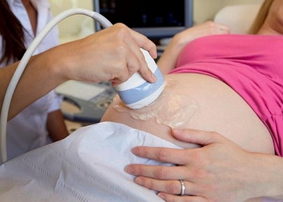
How is Ultrasound With Doppler Performed During Pregnancy?
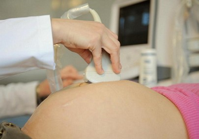
In most cases, especially if you are scanned in a paid clinic, Doppler ultrasound is no different from your scheduled ultrasound examination. The fact of the matter is that modern ultrasound machines are already equipped with the dopplerograph function. If the usual machine is slightly older than contemporary, then you will be directed for an examination in another place where such a device is available.
Conducting the scan using Doppler ultrasound during pregnancy is similar to the procedure of an ordinary ultrasound:
- You come into the office at the appointed time (no special preparation for that ultrasound is required);
- You lay on the couch and asked to lower the skirt or pants;
- Special gel is applied on the tummy and moving on the stomach with a special ultrasound "mouse", the doctor investigates the life inside you.
This is something that you will have to do when you will be directed to a Doppler. The doctor operates a little differently. He first on a normal mode of ultrasound explores the overall picture of the condition of the uterus and fetus. Then he determines the location of the vessel, which he also wants to look at more carefully (vessels in the umbilical cord, arteries of the brain and others). Then he switches on the function of dopplerograph. After that you can see the picture on the screen, which will show the state of the blood flow in the vessel. Then the device itself analyzes the received information and reports on deviations from the norm, if there are any. In general, the scan takes only a few minutes.
To Whom and When Ultrasound With Doppler is Prescribed During Pregnancy?
During gestation of the baby the doctor-gynecologist who is observing a pregnant woman, works out a definite plan of action. The development of this plan depends on the overall status of a woman, the presence of chronic diseases and addictions, fetal development, in accordance with the terms. But there are also some obligatory procedures, which are the same for every pregnant woman. One of such procedures is the scan with Doppler ultrasound.
This scan is done necessarily 2 times within 9 months, if there are no additional contraindications to it:
- On 22-24th week;
- On 30-34th week.
But if the doctor at the scheduled scan observes any abnormalities, then he has every right to send you to the Doppler velocimetry several times.
Additional indicators to conduct ultrasound with Doppler during pregnancy are:
- mother's high blood pressure;
- kidney problems;
- the presence of harmful habits-smoking-at the expectant mother;
- the existence of preeclampsia;
- multiple pregnancy;
- RH maternal-fetal conflict;
- pathology in previous pregnancies;
- differences in the size of the fetus, and terms of pregnancy;
- gestosis;
- presence of chronic and hereditary diseases (diabetes and so on);
- poor results of cardiotocography.
Is Doppler Harmful During Pregnancy?
The scan using ultrasound with Doppler is absolutely harmless and won't affect fetal development. Most likely, this scan is even useful. Why? Doppler is a very accurate scan of the fetus and its development. It happened so, that only by ultrasound with Doppler fetal hypoxia has been identified, and treatment was assigned in time that prevented an accident. Sometimes only Doppler ultrasound might see the entanglement of the baby with umbilical cord and its degree (double, triple, single, tight, not tight).
This is Doppler during pregnancy. Non of the scans during pregnancy should trouble a woman. Being nervous is bad for your health and the health of your unborn baby. Watch your habits, nutrition, behavior, and each new scan will delight you with the sounds of the heartbeat of the baby.